Conjuctivitis
The conjunctiva are the membranes that line the inside of the horse’s eyelids, and conjunctivitis means that these membranes have become inflamed. This may be a primary problem, but can also occur due to disease in other parts of the eye. Horses with corneal ulcers, for example, will often also have a degree of conjunctivitis. It is an extremely common condition, especially in mid-to-late summer, when flies play a big part in causing it.
Signs To Look Out For
- Swollen eyelids – the eyelids themselves will appear swollen. This is called chemosis. In mild cases this will only be visible on the inside of the lids, but when more severe, the eyelids can appear enlarged from outside as well.
- Discharge – varying amounts and types of discharge can be present. This can range from very watery to very thick and pus-like. In some cases the discharge is due to overproduction of tears (known as epiphora), in others then it is because the swelling has closed the tear duct, so tears cannot drain normally. In most cases both occur to some degree.
- Reddened eyelids – the inside of the eyelid may become diffusely red (called hyperaemia), or show an increased number of small blood vessels (called conjunctival injection). In some cases the white of the eye will also have an increased number of small blood vessels, known as scleral injection.
- Mild eye pain – the eye may be held slightly closed. Conjunctivitis is not usually associated with severe signs of pain.
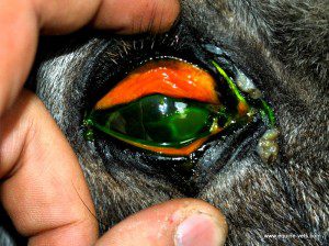
This horse has a corneal ulcer with secondary conjunctivitis. Notice how red the inside of the eyelids are, and also the increased amount of discharge at the corner of the eye.
How We Diagnose It
Conjunctivitis is often present to some degree with many different eye conditions, so it is important to determine if the signs we are seeing are primarily due to conjunctivitis, or whether they appear secondary to another problem. This relies on a thorough exam of the eye as described here.
The vet will also want to be certain that the tear duct (also known as the nasolacrimal duct) has not become blocked. They will do this by placing fluoroscein dye in the eye, and then watching for it to appear at the bottom of the duct, which lies just inside the nostril. This can take up to 20 minutes.
If the vet suspects an infectious cause, or if the conjunctivitis is not responding to treatment as expected, they may take a swab from the eye to culture bacteria, and alter antibiotic therapy based on this.
Treatment
Topical treatments are the usual starting point, and your vet may choose to use antibiotics, steroids, or a combination of the two. Antibiotics are used to deal with any bacterial infection, and steroids to reduce inflammation.
If your horse has a blocked tear duct, then this may need to be flushed with saline. Often eye drops will then be placed into the duct itself to help prevent it from becoming inflamed and becoming blocked again.
Flies can be an issue with these horses – they can cause the disease in the first instance, and even if not the initial cause, they will be attracted to the discharge and make the problem worse. If this is the case then a fly mask can be extremely useful. We have had good success using the “Guardian” style fly mask, which has the added advantage of protecting from UV light.
Outcome
The prognosis for horses with conjunctivitis is excellent, and most will resolve quickly with no lasting effects. On very rare occasions the tear duct may become scarred, and no longer be able to effectively drain tears away from the eye. This may lead to a persistently weepy eye.