Laminitis
Laminitis is one of the most common causes of lameness in horses, and especially ponies in the UK. The lameness can vary from a mild alteration in gait at walk, right through to refusal to move, and even recumbency (lying down). The majority of cases can be treated effectively, with a full recovery, but in severe cases, or those that are not treated quickly and effectively, laminitis can be a life threatening condition. There is a great deal of research currently being carried out into laminitis, and new therapies have become available in the last few years, however, there is a lot that is still unknown. In order to understand laminitis, we must first look at the structure and function of the horse’s foot.
The Horse’s Foot
An average horse weighs 500kg (1100 pounds), and carries 60% of its weight on its forelimbs, 40% on its hindlimbs. This means that around 150kg is being supported by each fore foot, and 100kg by each hind foot. The majority of this weight lies in the horses thorax and abdomen, and is supported by the bony column of each limb. The weight is then transferred from the bony column of the limb into the hoof wall, to be supported by the foot. This transfer of weight from limb to foot occurs exclusively between the pedal bone and the hoof wall, via connections called the laminae. The entire weight of horse is transferred through these 4 collections of laminae, one in each foot, and they must therefore be tremendously strong.
The role of the laminae is two-fold: firstly, as described above, to act to transfer weight from limb to foot. Secondly, they act as the attachment from pedal bone to foot that means movement of the pedal bone will result in movement of the foot.
What Is Laminitis?
In veterinary (and medical) terminology, an ‘–itis’ means inflammation of an area, for example, hepatitis (inflammation of the liver) or tendonitis (inflammation of a tendon). Similarly, laminitis simply refers to inflammation of the laminae within the foot. Inflammation is defined by 5 key characteristics, and we will look at how these relate to laminitis:
- Heat – this is commonly felt as an increased temperature of the dorsal (front) hoof wall
- Pain – seen as alterations in gait, reluctance to move, recumbency
- Swelling – although not seen, this has a key relevance to the condition
- Redness – again, not often seen, although red horn at foot trimming can reflect previous inflammation
- Loss of function – both weight supporting and foot movement functions can be impeded
It is these signs of inflammation of the laminae that will guide us in making a diagnosis of laminitis. Unfortunately the overall picture is not that simple – the 5 categories seen above show us that there is laminar inflammation, and this is defined as acute laminitis. There is also a condition known as chronic laminitis, and a horse may have acute or chronic laminitis, and in some instances both.
Chronic laminitis is defined as an alteration of the internal anatomy of the foot (although it will often lead to changes in the external anatomy as well), that occurs as a result of acute laminitis. These changes are broadly divided in two:
- Pedal bone rotation (“founder”) – the pedal bone rotates, compared with either the hoof wall (capsular rotation), or the pastern bones (phalangeal rotation)
- Pedal bone sinkage (“sinker”) – the pedal bone drops vertically downwards within the foot
Again, these two forms of chronic laminitis can occur alone or in combination. Looking back at our 5th sign of inflammation (loss of function), we can explain why these anatomical changes occur. Rotation occurs due to the impairment of the ability of the laminae to translate movement of the pedal bone into movement of the hoof. Force applied to the pedal bone by the deep digital flexor tendon (DDFT) therefore moves the bone within the hoof, rather than the bone and hoof together. Sinking occurs due to the impairment of the ability of the laminae to transmit weight from limb to hoof capsule, with subsequent distal (downward) movement of the pedal bone. The pedal bone and hoof wall shear apart rather than stay firmly attached.
What Causes Laminitis?
Laminitis is a disease that can occur due to a large number of different reasons, and in individual cases there may be more than one cause. We strive to find the underlying reason(s) in all cases, as it can greatly assist in treatment, and are successful in finding the cause in the great majority.
- Endocrine induced laminitis
- This is by far and away the most important aspect of laminitis and contributes the largest cause.
- The endocrine causes have recently been recognised to be extremely important, and any horse with laminitis should be tested routinely.
- There is more information in our fact sheets on Cushing’s Disease and Equine Metabolic Syndrome.
- Pasture induced laminitis
- This is the “classic” form of laminitis with which we are likely most familiar.
- It typically occurs in Spring/Summer, when horses eat a large amount of lush grass.
- A similar situation occurs in horses that raid feed bins!
- This sudden dietary change disrupts the large intestine, and allows absorption of vaso-active compounds (substances that affect blood vessels).
- This affects the blood supply to the foot, resulting in too little oxygen reaching the laminae (laminar hypoxia).
- The alteration may be to close arteries to the laminae, or may be to open arterio-venous shunts (vessels that bypass the laminae). More research is currently being performed on this.
- It is thought that most cases of pasture-associated laminitis occur because of an underlying metabolic problem such as “endocrine or endocrinopathic laminitis”
- Weightbearing laminitis
- A severe injury to one leg, that results in massively increased weight bearing in the opposite foot can trigger laminitis.
- Any horse with prolonged severe lameness to one leg should have preventative measures taken on the opposing foot.
- Endotoxaemia induce laminitis
- Endotoxaemia is a medical condition that results in bacterial components entering the bloodstream, which can trigger laminitis.
- It can be caused by retained placental membranes (RPM) where a mare retains her afterbirth after foaling, severe colitis (inflammation of the large intestine) and some severe forms of colic.
- Corticosteroid Induced Laminitis
- These cases commonly have underlying endocrine problems (EMS/Cushings) that increases the risk of laminitis.
- There are numerous reports of steroid administration triggering laminitis, through an unknown mechanism. There are some similarities with Cushing’s Disease induced laminitis.
- Although the incidence is low, the consequences can be severe, so before administering steroids your vet will always talk to you about the risks and benefits of using steroids.
Clinical Signs of Laminitis
We have explored in some depth the changes that occur in laminitis, but more importantly we must also consider how these changes are reflected in the horse.
Acute laminitis has some typical signs that are frequently seen, and we will list them and discuss why they occur:
- Increased Digital Pulses
- Two arteries course down the back of the leg, and in acute laminitis can usually be felt to be pulsing. This compares with a normal horse, where the pulses can just barely be felt.
- The easiest site to feel these pulses are just above the fetlock (close to the windgall), and either side of the back of the pastern.
- In order to understand why these pulses increase, we must first understand what a pulse actually is. We all know that a beat of a pulse represents the beat of the heart, but in fact a pulse has two components – the peak of a pulse is the blood pressure when the heart beats (systolic B.P.), and the ebb of the pulse is the blood pressure when the heart relaxes (diastolic B.P.). It is the difference between the two pressures that is felt as a pulse. Therefore, an increase in the beat of a pulse can either be from an increased systolic B.P., or from a decreased diastolic B.P.. In laminitis it is this decreased diastolic blood pressure that creates the bigger pulse, and this can occur for two reasons.
- When they become inflamed, blood vessels become more leaky, so more blood leaves the arteries in the feet, lowering the diastolic blood pressure.
- Arterio-venous (AV) shunts are blood vessels that divert blood straight from artery to vein, avoiding the capillary bed of the foot. If these shunts open then blood will leave the artery extremely quickly, again lowering diastolic blood pressure. There is more on these AV shunts later.
- Warm hoof walls
- Increased temperature of both the hoof wall and the coronary band can often be felt in horses with laminitis, which occurs for two reasons:
- Inflammation – as we saw earlier, one of the key signs of inflammation is heat
- Increased blood flow – more blood flowing through the foot will result in an increased temperature
- This is a sign that must be interpreted with some caution – recent research has shown that normal horses do not supply blood to their legs all the time, but in fact do so intermittently. If we happen to feel the hoof wall during one of these intermittent flushes of blood supply, then the foot will feel warmer than normal with no abnormality present.
- Increased temperature of both the hoof wall and the coronary band can often be felt in horses with laminitis, which occurs for two reasons:
- Lameness
- Lameness can vary greatly, but is often graded using a scale known as Obel Grading:
- Grade I – alternate lifting of feet, no lameness at walk
- Grade II – stiffness at walk, resists turning, lame at trot
- Grade III – lame at walk, resists lifting feet
- Grade IV – reluctant to move unless forced
- Gait alterations are also seen, the most common being a heel-toe gait. The greatest number of laminae are at the toe of the foot, so this is where the greatest pain is felt. The horse therefore lands on its heels to reduce the pain felt.
- Lameness can vary greatly, but is often graded using a scale known as Obel Grading:
- Hoof tester Response
- Your vet will apply hooftesters to your horse’s foot, and pain is often elicited just in front of the frog.
- This is not always present, and can be widely affected by the depth of the horse’s sole, and its hardness.
- Altered Stance
- The typical “laminitic stance” is a horse leaning backwards, with its front legs stretched out far in front. This occurs because of the increased weight borne through the fore limbs, so the horse is trying to support more weight on its back end.
- While extremely well known, this stance is actually not seen all that often in cases of laminitis.
Chronic laminitis has less reliable clinical signs, although is often also accompanied by some degree of acute laminitis.
- Alterations in hoof growth
- Concave dorsal hoof wall surface
- Hoof wall rings that converge toward the toe
- Both these changes occur due to faster hoof wall growth at the heel than at the toe
- External indicators of sinkage
- Depression over dorsal coronary band
- Convex sole
- Widened white line
- This commonly occurs if there has been rotation of the pedal bone
- Variable lameness
- Variable response to hooftesters
Treating Acute Laminitis
The majority of horses that we see with laminitis are suffering with the acute form, and so we will discuss treatment of this initially. It is uncommon for us to see a horse with chronic laminitis that has not suffered with a noticeable episode of acute laminitis previously.
We have four major aims in treating acute laminitis; pain management, restoration of blood flow, support of the foot and removal of underlying cause. All of the measures that we take are trying to achieve one or more of these aims.
- Strict box rest
- This is vitally important for several reasons
- Movement puts unnecessary forces through already weakened laminar bonds, causing further inflammation and instability.
- Restricting movement lessens the risk of pedal bone sinkage or rotation.
- Movement is painful, so we must restrict it for welfare reasons.
- Deep bedding
- A softer bed reduces concussion on the foot, and relieves pain.
- Restricted Diet
- Horses on box rest have far lower energy needs.
- Many cases of laminitis are associated with weight problems, and restricting diet is appropriate for these horses.
- Pasture-induced laminitis (see later) requires a low carbohydrate diet.
- Soaked hay, with a low calorie hard feed is commonly utilised.
- Frog Support
- In many cases, supporting the frog is appropriate.
- This takes pressure away from the painful sole, and reduces tension in the DDFT. Reduced DDFT tension can help prevent pedal bone rotation.
- Many varieties are used, including hoof putty, rolled up bandages and styrofoam pads.
- Anti-inflammatory Drugs
- Non-steroidal Anti-Inflammatories (NSAIDs) are commonly used.
- These drugs reduce inflammation and control pain.
- The two most frequently used are phenylbutazone (“Bute”) and flunixin (Finadyne).
- In terms of their ability to reduce pain, there is no evidence to show any difference, although traditionally Bute has been regarded as being more effective for orthopaedic conditions.
- Flunixin protects against the cardiovascular effects of endotoxaemia, by preventing blood vessel dilation. This can be useful in closing AV shunts (see later).
- Flunixin can be given for a maximum of 5 days, so after this period bute is commonly used.
- Vasodilating Agents
- The commonly used drugs are peripheral vasodilators, the most common being acepromazine (ACP, Sedalin).
- The aim is to open up constricted veins and improve blood supply to the laminae.
- ACP also has a mild tranquillizer effect that can be beneficial in reducing movement.
- Glyceryl trinitrate (Percutol), a topical cream with vasodilator, anti-inflammatory, analgesic and anti-free radical effects used to be in common usage. Unfortunately, this product is no longer manufactured.
Unfortunately in some cases, even with prompt and appropriate treatment, some changes in the position of the pedal bone occur, and the acute laminitis progresses to chronic laminitis.
Treating Chronic Laminitis
Chronic laminitis is a challenging, and sometimes extremely frustrating condition to treat. What works for one horse will not necessarily work for another, even if all the circumstances appear the same. We suspect that there may be chronic laminitic changes in horses that have suffered an attack of acute laminitis that has not resolved swiftly (within approximately one week), and if this is the case then we will investigate further.
Chronic laminitis is primarily a radiographic diagnosis, meaning that it is made based on x-ray findings. Radiographs (x-rays) will reveal the exact position of the pedal bone within the foot, relative to the pastern bones and to the hoof wall. They will also reveal any sinking relative to the coronary band.
Solar depth can be assessed, which is extremely useful to know when it comes to corrective trimming and shoeing.
In long standing chronic laminitis cases, there may be remodelling of the pedal bone. The dorsal (front) surface of the bone may take on a curved concave appearance, and the tip of the bone may start to become re-absorbed.
The basis of treatment of chronic laminitis is corrective farriery, and your vet will work closely with your farrier at all stages. We are fortunate to have a mobile digital x-ray machine, which allows us to take radiographs and see results instantly. We can meet your farrier at your stables, and take x-rays before, during and after trimming and shoeing to ensure the best result possible.
As stated previously, no two horses with chronic laminitis are the same, but the starting point of treatment is often similar. Heart-bar shoes are complete round shoes, with a triangular extension over the frog, which are the usual first shoe that is tried. In horses with thin soles then sole filler may also be used for protection.
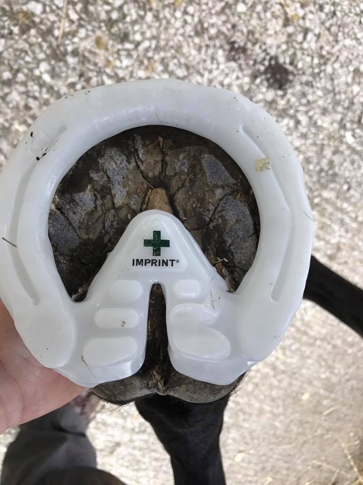
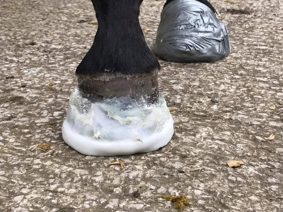
Imprint Shoes and Filler
In certain cases heel wedges may be appropriate.
Heart Bar Shoe
Dorsal hoof wall resection (DHW) is a procedure where a small piece of the front hoof wall is removed, which can have several benefits.
When the pedal bone rotates, the space it vacates is filled with fluid, which can exert pressure within the foot. A DHW relieves this pressure, and can have superb results. Removal of this pressure also aids in restoring blood flow to the laminae. The DHW can also help in promoting proper regrowth of hoof wall, by allowing the hoof wall laminae and the pedal bone laminae to re-align.
Dorsal Wall Resection
Prognosis
Accurately predicting an outcome for a horse with laminitis is extremely difficult. One recent study found that the only reliable indicator of prognosis was how well the horse responded to initial treatment. This is something of a circular argument and not very useful to us in giving a prognosis for your horse. In general, a horse that has a single bout of acute laminitis, and recovers quickly, has an extremely good chance of returning to its previous level of work.
Horses with underlying conditions such as Cushing’s Disease and Equine Metabolic Syndrome are more difficult to manage, and have a poorer prognosis. However, we have many horses currently being treated for these conditions and doing extremely well.
The Obel grade of lameness at presentation (see above) also has some relevance, with more severely affected animals taking longer to recover, and not responding as well.
Your vet will talk through all these factors if your horse suffers from laminitis, and try and give you the best idea possible of how well treatment will progress.